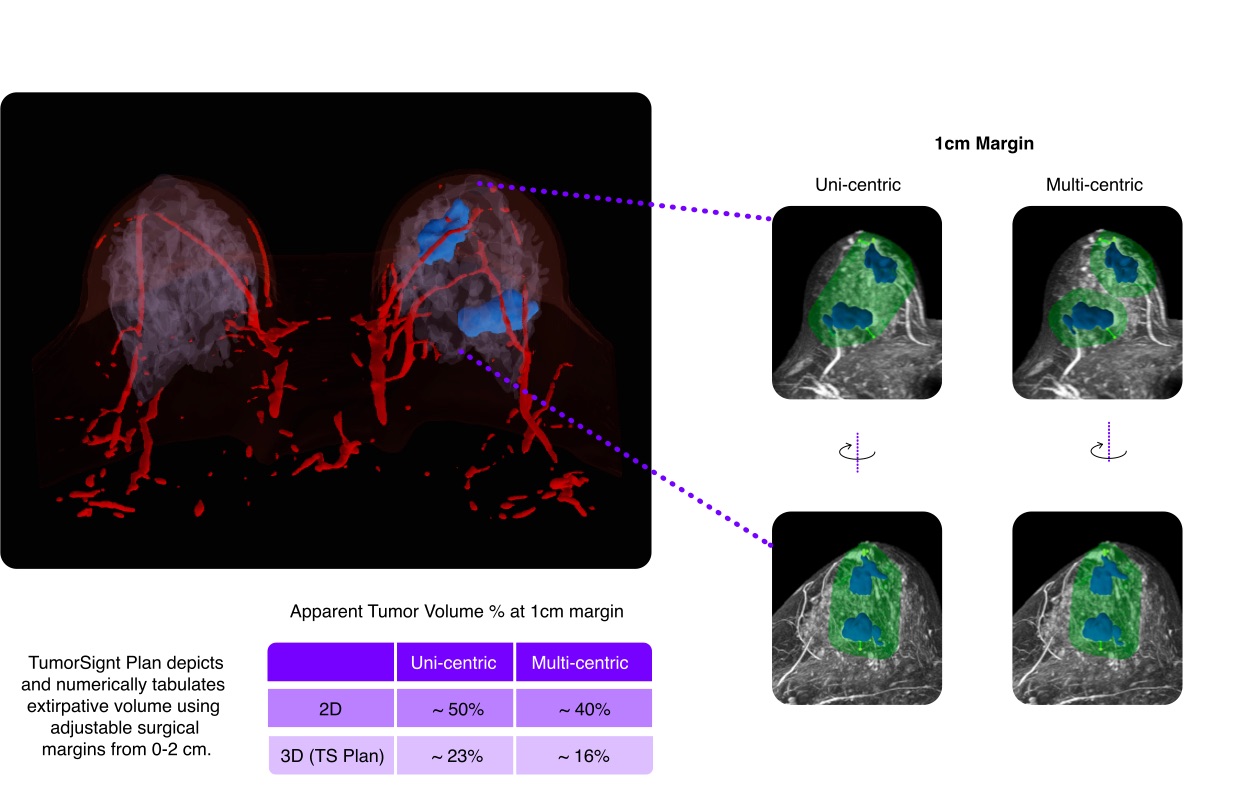
TumorSight Viz provides surgical oncologists with 3D spatial visualizations of breast cancer using standard-of-care medical imaging (DCE MRI) and critical measurements that are informative prior to surgical planning (e.g., Breast Conservation Surgery). Clear 3D “digital twin” renderings automatically display the tumor in the context of auto-segmented anatomical structures (skin, vessels, chest, fat, gland).
Automatic segmentation (artificial intelligence) and displays tumor shape, size, morphology, and location.
Reliable volume calculations to inform pre-surgical decision-making to assess breast-conserving surgery or mastectomy options.
Plan optimal surgical outcomes in breast-conserving surgery using measurements to key anatomical features.
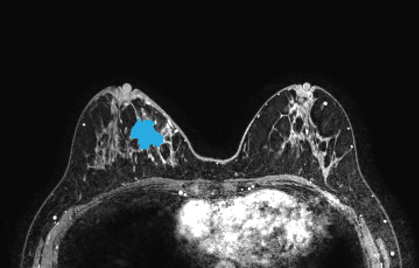
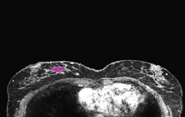
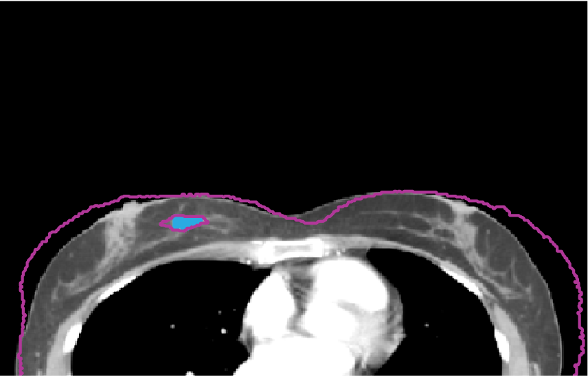
SimBioSys has developed a research-use application that is designed to convert an MRI from a prone to supine operative position, enabling enhanced surgical & reconstructive visualization. Supine models in The novel this research use application will be trained to incorporate the effects of gravity on different tissues, movement of tissues relative to adjacent structures, and variability in skin elasticity as a function of patient demographics.
The images below show the standard MRI in the prone position, the research use application’s conversion to the supine position, and the CT in the supine position (tumor in blue) overlayed with the application’s supine conversion outlined in purple.
*This feature is currently approved for research use only

The 3D model and measurements provided by TumorSight Viz & CDS gives breast surgeons additional information to consider breast conservation surgical options which can minimize surgery severity and improve patient quality of life. Additionally, the 3D model’s 360° depiction of the tumor and surrounding tissues are designed to enhance collaborative decision-making between the surgeon and patient.
When surveyed, 70% of surgeons rated the tool as valuable or very valuable overall. They noted the greatest value when used in specific cases, such as multi-focal and multi-centric, DCIS, large tumors, and tumors close to the skin or nipple.
Source: Internal survey
TumorSight Viz uses individual patient standard-of-care medical imaging and diagnostic data, if available, as inputs. The software identifies tumor tissue automatically with trained AI and creates a 3D tumor along with surrounding tissue to calculate breast and tumor volume along with distances to nearby anatomical features.
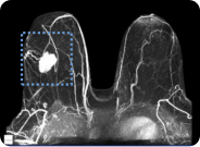
Dynamic Contrast-Enhanced MRI

Convolutional Neural Networks
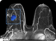
Patient-specific info & analytics